The pathogenesis of melasma is important when considering
laser treatment. Kim et al1 reported increased vascularity as
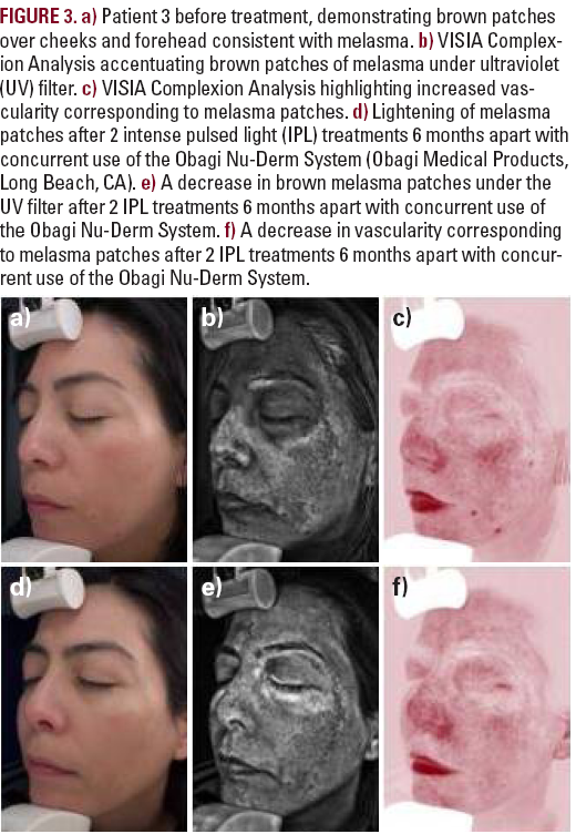
a major finding in melasma with increased amounts of VEGF
and blood vessels within melasma lesional skin. It has been
postulated that ultraviolet (UV) radiation-induced dermal
inflammation activates fibroblasts and stem cell factors in
melasma dermal skin, causing melanogenesis. The increased
vascularity could be why melasma occurs in select regions and
not uniformly across the face, despite equal UV damage.
Vascular endothelial growth factor has also been shown to stimulate
the release of arachidonic acid, and the metabolites of this
pathway may affect melanogenesis. Steroids in triple-agent
creams used to treat melasma can also induce telangiectasias,
possibly exacerbating this component of melasma.1 Bak et al8
reported increased nerve growth factor and neural endopeptidase
in melasma lesional skin, also suggesting its association
in the pathogenesis of melasma.
Multiple studies have shown varied effectiveness of lasers in
the treatment of melasma. The nonablative 1,550-nm fractional
laser has been used to treat melasma with greater patient
satisfaction 3 weeks after treatment compared with topical triple-agent therapy of HQ 5%, tretinoin 0.05%, and triamcinolone
acetonide 0.1% cream. It was thought the laser brought greater
satisfaction early on because of a faster initial clearance and a
possible increased effectiveness in treating dermal melasma.
However, 6 months posttreatment, the pigmentation returned
in both treatment groups with equal patient satisfaction rates.
Side effects of the 1,550-nm laser treatment included erythema,
burning sensation, edema, and pain. Kroon et al6 noted no
postinflammatory hyperpigmentation (PIH) with this treatment
modality using conservative settings.
Wind et al9 described the use of the nonablative 1,550-nm fractional
laser in the treatment of melasma, and 9 patients (31%)
developed PIH after 2 or more laser sessions. The increased PIH
seen in the study is likely secondary to more aggressive treatment
settings. Skin findings and side effects associated with
topical triple-agent therapy included erythema, scale, and burning.
Triple-agent topical therapy is still the treatment of choice
because of similar efficacies 6 months posttreatment.10-11
The Q-switched Nd:YAG laser has also been reported to temporarily
improve melasma with common complications, including
hypopigmentation, melasma recurrence, and rebound hyperpigmentation.
Transient erythema, transient burning, and slight
edema occurred for 1 hour postprocedure. Wattanakrai et al12
found decreased epidermal and dermal pigmentation for up to
1 year after 10 weekly treatments with the Q-switched Nd:YAG
laser at subthreshold photothermolytic fluencies (<5 J/cm2).
However, rebound hyperpigmentation was common, and the
risk of mottled hypopigmented macules increased with greater
number of laser sessions.12,13 Narrowband UVB has successfully
been used to treat depigmentation with good clinical results.13
The short-pulsed deep Er:YAG laser temporarily but effectively
reduces epidermal type melasma, with a recurrence upon discontinuation
of treatment.14
A review of the literature suggests that laser and light source
treatments can result in rebound hyperpigmentation, relapse,
and darkening of melasma. It has been postulated that the laser
unmasks a previous subclinical melasma. This is thought to
be secondary to stimulation of hyperactive melanocytes, which
can increase melanin production and therefore pigmentation.15
Intense pulsed light is a noncoherent filtered flashlamp light
source, emitting light between 515 and 1,200 nm. Filters allow
for selective photothermolysis of chromophores, including
melanin and hemoglobin.5,16 Since its introduction in 1992,
there are now more than 20 different IPL devices available
worldwide.17 Each IPL device has a unique set of wavelengths,
fluences, pulse durations, epidermal temperature effects, and
other pulse parameters in addition to duration, such as unifor-

