 image analysis of LifeViz App (QuantifiCare, France) and then compared to QuantifiCare’s reference population database of normal aging skin, adjusted for age, sex, and skin type; the 3-dimensional high-resolution micro-imaging of skin surface of those participants were taken using micro imaging system (QuantifiCare, France).
 Griffiths scale
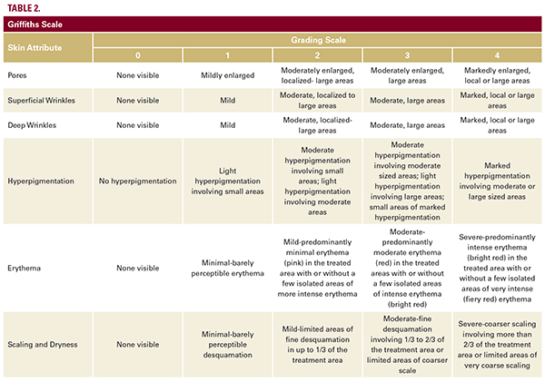
We have used the original scale described by Griffiths.22 Specific skin attributes are shown in Table 2.
Statistical Methods
All statistical analysis was performed using R (Ver. 3.4.1). Significance was set a priori at PTwo-sample student’s t-test was used to compare the improvement at 6 weeks and 12 weeks between the two groups (PG vs FFG) for each of the parameters for analysis of the data acquired using Cortex and Quanti Care systems, and histopathology (epidermal thickness) and immunohistopathology (Ki67) data. Descriptive summaries are expressed as means +/- standard deviation (SD). Fisher’s Exact test was used to compare the improvement percentage at 6 weeks and 12 weeks between the two groups (PG vs FFG) to analyze the Griffiths scale data. Descriptive summaries are expressed as frequency and percentage.
RESULTS
The histopathological analysis revealed an increase of the epidermal thickness in all four participants of the FFG assigned for skin biopsy and less or no increase in the PG. The increase in epidermal thickness in the FFG was 0.09±0.02 mm (average) vs 0.02±0.03 mm in the PG (P=0.027; Figure 4A, B, C). The regimen did not appear to cause inflammation, as was seen in the histopathology analysis via lack of inflammatory cells and absence of spongiosis in FFG (also none seen in PG; Figure 4D). The microscopic lack of inflammation was

