trichrome in order to visualize wound morphology and collagen
deposition, respectively.
Cytokine Analysis
Mice were sacrificed on days 3 and 7 for cytokine analysis in all groups. Tissue was flash-frozen using liquid nitrogen, homogenized
in ice-cold phosphate buffered saline (PBS) with Protease Inhibitor Cocktail (Sigma-Aldrich, St. Louis, MO). Samples were centrifuged at 10,000 G for 20 minutes to remove debris. Aliquots of the supernatants were assayed for total protein content by the BCA method. Cytokine analysis was carried out using BD Cytometric
Bead Array (CBA) Mouse Th1/Th2/Th17 Cytokine Kit (BD Biosciences, San Jose, CA) according to the instruction manual. Samples were examined using BD FACS Canto II flow cytometer at 485 and 633nm and results were analyzed with Flowjo software.
RESULTS
Clinical Wound Closure
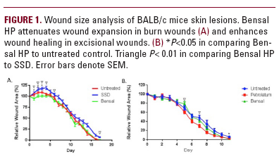
Topical Bensal HP applied to burns (Figure 1a) inhibited wound expansion during the first six days post-injury with significant differences over the untreated group on days 1-3, and over SSD on days 1-4,6, and 17. The majority of wounds in both the untreated
and Bensal HP-treated mice reached closure by day 17. In excisional wounds (Figure 1b), Bensal HP showed a significant improvement in wound healing over the untreated group by the final day of the study period, with petrolatum significantly outperforming
Bensal HP on days 5 and 7 post-injury. Wounds in all three study groups reached closure by day 11 post-injury.
Histology
H&E staining of burn wounds on day 7 (Figure 2a) revealed a serous neutrophil rich crust overlying a homogenized inflammatory dermis/granulation in both control and bensal