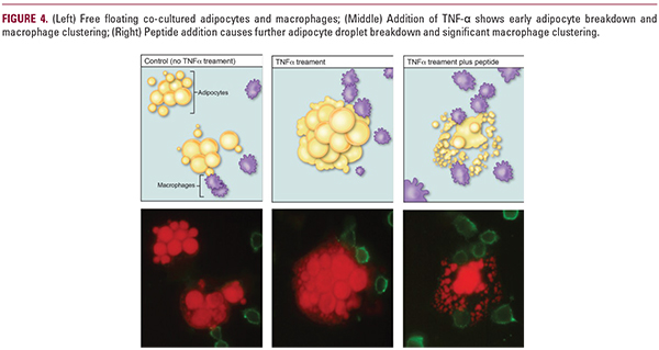
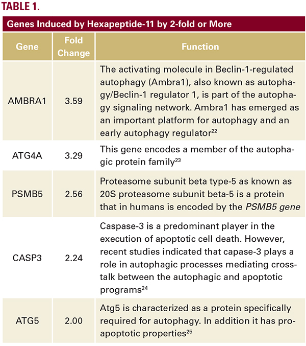
 mix and treated and untreated cells were analyzed by real time PCR analysis at 72 hours.Results: 72-hour treatment with hexpeptide 11 increased expression of 5 major autophagic genes (AMBRA1, ATG4A, PSMB5, CASP3, and ATG5) by 2-fold or greater (Table 1). Next, an in vitro model was designed to assess efficacy of the peptide with lipid droplet breakdown and macrophage clustering. 3T3-L1 adipocytes were cultured and developed; macrophages were then added to the adipocytes. They were then exposed to TNF-alpha (~25nM) for 24 hours, which caused apoptosis in approximately 70% of adipocyte cells. This provides a perfect model for adipose apoptosis as seen in non-surgical fat reduction. One set of macrophage adipocyte co-culture was also exposed to the hexapeptide and adipose droplet breakdown and macrophage clustering was observed in each group. Macrophages and adipocytes were stained to facilitate identification. The experiment was completed in triplicate.Results (Figure 4): Co-culturing of macrophage and adipocytes revealed free floating cells with little macrophage clustering.The addition of TNF-α increased droplet breakdown and macrophage clustering. The most significant adipose breakdown and macrophage clustering was observed following the addition of the peptide.The gene expression studies and co-culture model thus provide encouraging in vitro validation of peptide efficacy. Targetingof the hair follicle point of entry for the peptide is achieved by combining the peptide with a liposome designed specifically for follicular penetration (particle size of liposome vesicles ~185nm) as described above, ensuring entry to the dWAT compartment.