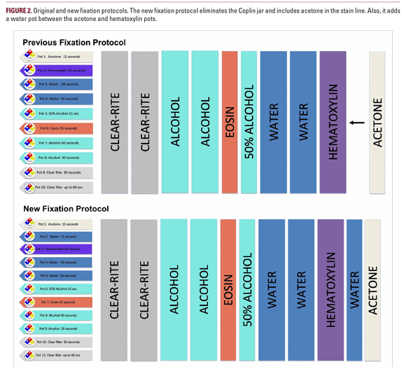
caused by weak staining, possibly related to an interaction between hematoxylin dye solution, which is water-based, and acetone. Our protocol for the Linistain (SLS) stain line (Thermo Scientific Linistat Linear Stainer) included dipping the slide into a Coplin jar of acetone for ~15 seconds and then dipping it directly into hematoxylin on the SLS stain line (Figure 2). This protocol using acetone in a Coplin jar rather than on the stain line was the traditional protocol at our MMS laboratories at the time. We adjusted the SLS stain line protocol by adding a 15 second water rinse between the acetone and hematoxylin pots (Figure 2). Also, we eliminated the Coplin jar and placed the acetone on the SLS stain line. In order to validate our new fixation technique, we tested the same specimen tissue and compared the old fixation protocol with our new fixation protocol.
Internal sections cut at 4-5 μm thickness were standardized for our assay. The first section was stained with the new fixation protocol and the second section was stained with the original fixation protocol. Both slides moved through the entire SLS stain line, were cover-slipped, and viewed microscopically by separate MMS surgeons and dermatopathologists to validate the quality of slide preparation. Sixty slides were run through this assay, 35 through the new fixation protocol and 25 through the previous fixation protocol. Of the 25 slides that were test-

