Plaque Sarcoidosis
Plaque sarcoidosis is characterized by elevated lesions >5 mm in size.6 Plaques can be found on the face, extremities, or trunk, and may occur alone or in multiples. When plaques present in multiples, they are typically seen in a symmetric distribution (Figure 2).5 Plaques are more likely to develop in deeper skin layers than papules.6
Differential diagnoses: Lichen planus, Psoriasis, Cutaneous T-cell lymphoma.
Differential diagnoses: Lichen planus, Psoriasis, Cutaneous T-cell lymphoma.

Scar Sarcoidosis
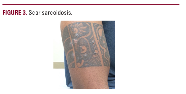
Scar sarcoidosis involves patches that appear in areas of previous scarring. The patches may present as erythematous or violaceous in color and will affect areas such as the face, trunk, scalp, and extremities. The initial scarring can be caused by any mechanical trauma to the skin including venipunctures, previous infections, and tattoos (Figure 3). These lesions themselves are often asymptomatic and can be an indication of a sarcoidosis exacerbation.6
Differential diagnoses: Keloids, Hypertrophic scar.
Differential diagnoses: Keloids, Hypertrophic scar.
Lupus Pernio
Lupus pernio more commonly affects women with skin of color.6,9 It presents as indurated papules or plaques that vary in color from red to purple.9 Lupus pernio is seen predominantly on the skin over the cheeks, nose, lips, and ears.6
Differential diagnoses: Lupus erythematosus, Lupus vulgaris, Leprosy.
Differential diagnoses: Lupus erythematosus, Lupus vulgaris, Leprosy.
Nodular (Subcutaneous) Sarcoidosis
Nodular sarcoidosis, also known as Darier-Roussy sarcoidosis, involves non-tender firm subcutaneous nodules that are mobile and 0.5 - 2 cm in size (Figure 4).6
Differential diagnoses: Granuloma annulare, Lipomas.
Differential diagnoses: Granuloma annulare, Lipomas.

Ulcerative Sarcoidosis
Ulcerative sarcoidosis may arise with or without the presence of a pre-existing lesion on the lower extremities.3 Ulcerative lesions are twice as likely to develop in women and individuals with darker skin tones.6
Differential diagnoses: Ulceration from stasis dermatitis, Cutaneous tuberculosis.
Hypopigmented Sarcoidosis
Hypopigmented sarcoidosis typically presents in patients with darker skin tones as well-demarcated hypopigmented macules and can also present as papules or nodules.6 The papules are erythematous or skin-colored and may develop at the center of a hypopigmented lesion, giving the appearance of a fried egg.8
Differential diagnoses: Seborrheic dermatitis, Pityriasis alba, Vitiligo.
Ichthyosiform Sarcoidosis
Ichthyosiform sarcoidosis is rare and presents as scaly hyperpigmented plaques that are polygonal in shape and vary in color from gray to brown.8 These plaques are commonly found on the lower extremities and are nontender and nonpruritic.6 Approximately 95% of patients with ichthyosiform sarcoidosis will develop systemic sarcoidosis.6
Differential diagnoses: Eczema, Ichthyosis vulgaris.
Nonspecific Skin Findings
Erythema Nodosum
Erythema nodosum is caused by inflammation of subcutaneous fat (panniculitis) and is characterized as tender erythematous nodules that typically present on the shins anteriorly. It more commonly presents in patients of European, Puerto Rican, and Mexican descent, and often remits without treatment.6 Erythema

