To the Editor,
The nose is central to facial symmetry and appearance. Reconstruction of defects spanning the lower nasal subunits poses particular challenges, given the need to preserve free margins, three-dimensional anatomy of the nares, and airway patency.1,2
Nasal defects frequently span multiple subunits and require complex, locoregionally tailored reconstruction.2,3 Flaps offer broad applicability, robust vascularity, and superior aesthetic outcomes compared with full-thickness skin grafts (FTSGs), but at the cost of greater invasiveness and risks including pincushioning, necrosis, and contour irregularities.1,4 FTSGs and composite or batten grafts offer a less invasive, singlestage option. However, limitations include suboptimal texture match, alar notching, and inferior cosmesis.1,5 Primary closure offers simplicity, efficiency, minimal invasiveness, excellent tissue match, and smaller scars when utilized appropriately but is traditionally avoided in lower nasal subunits due to concern for distorting free margins.1
In this study, we aim to challenge this paradigm by highlighting scenarios in which primary closure can offer minimal invasiveness yet excellent cosmesis.
Surgical Technique
Assessment begins with evaluating defect size and tissue mobility. In this series, successful closures were achieved in small- to medium-sized defects extending most commonly to adipose tissue, reflecting clinical judgement rather than formal guidelines. Tissue mobility was assessed by placing one finger intranasally and one externally, then gently pinching the defect horizontally along the alar curvature to determine whether closure could be achieved without excessive tension or nare distortion.
If appropriate, guiding sutures are placed at the periphery in the mid-deep dermis to verify that closure with a rounded contour matching the contralateral alar rim is achievable. Once rim preservation is ensured, standing cones are excised, and closure is completed in a linear or curvilinear fashion from edge to center using simple interrupted 6-0 nylon sutures, typically without deep dermal sutures. Delaying cone excision preserves the option to abort primary closure if tension proves excessive. Given the lack of deep dermal sutures and high tension along the alar rim, suture removal is prolonged to 2 to 3 weeks postoperatively to allow greater tensile strength development. Cosmetic outcomes were evaluated by the performing surgeon using dermoscopy and qualitative patient-reported outcomes.
Case Series
Nine patients underwent primary closure following Mohs micrographic surgery (MMS) excision of non-melanoma skin cancer of the nasal subunits. The median number of MMS stages to clear margins was one (range: 1-2). Defects spanned the ala (n=5, 55.6%), columella (n=2, 22.2%), infratip (n=1, 11.1%), and soft triangle (n=1, 11.1%). Seven defects (77.8%) had depth limited to adipose tissue, with two (22.2%) extending to cartilage. Mean post-excision longest dimension and area were 9.00 ± 2.16 mm and 52.9 ± 21.2 mm2, respectively. Mean followup was 236 ± 175 days.
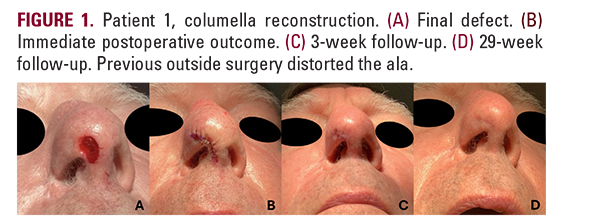
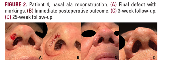
Clinical images for two patients are shown in Figures 1 and 2. Cohort characteristics and additional images are provided in Supplementary Tables and Figures (Mendeley Data Link).
The nose is central to facial symmetry and appearance. Reconstruction of defects spanning the lower nasal subunits poses particular challenges, given the need to preserve free margins, three-dimensional anatomy of the nares, and airway patency.1,2
Nasal defects frequently span multiple subunits and require complex, locoregionally tailored reconstruction.2,3 Flaps offer broad applicability, robust vascularity, and superior aesthetic outcomes compared with full-thickness skin grafts (FTSGs), but at the cost of greater invasiveness and risks including pincushioning, necrosis, and contour irregularities.1,4 FTSGs and composite or batten grafts offer a less invasive, singlestage option. However, limitations include suboptimal texture match, alar notching, and inferior cosmesis.1,5 Primary closure offers simplicity, efficiency, minimal invasiveness, excellent tissue match, and smaller scars when utilized appropriately but is traditionally avoided in lower nasal subunits due to concern for distorting free margins.1
In this study, we aim to challenge this paradigm by highlighting scenarios in which primary closure can offer minimal invasiveness yet excellent cosmesis.
Surgical Technique
Assessment begins with evaluating defect size and tissue mobility. In this series, successful closures were achieved in small- to medium-sized defects extending most commonly to adipose tissue, reflecting clinical judgement rather than formal guidelines. Tissue mobility was assessed by placing one finger intranasally and one externally, then gently pinching the defect horizontally along the alar curvature to determine whether closure could be achieved without excessive tension or nare distortion.
If appropriate, guiding sutures are placed at the periphery in the mid-deep dermis to verify that closure with a rounded contour matching the contralateral alar rim is achievable. Once rim preservation is ensured, standing cones are excised, and closure is completed in a linear or curvilinear fashion from edge to center using simple interrupted 6-0 nylon sutures, typically without deep dermal sutures. Delaying cone excision preserves the option to abort primary closure if tension proves excessive. Given the lack of deep dermal sutures and high tension along the alar rim, suture removal is prolonged to 2 to 3 weeks postoperatively to allow greater tensile strength development. Cosmetic outcomes were evaluated by the performing surgeon using dermoscopy and qualitative patient-reported outcomes.
Case Series
Nine patients underwent primary closure following Mohs micrographic surgery (MMS) excision of non-melanoma skin cancer of the nasal subunits. The median number of MMS stages to clear margins was one (range: 1-2). Defects spanned the ala (n=5, 55.6%), columella (n=2, 22.2%), infratip (n=1, 11.1%), and soft triangle (n=1, 11.1%). Seven defects (77.8%) had depth limited to adipose tissue, with two (22.2%) extending to cartilage. Mean post-excision longest dimension and area were 9.00 ± 2.16 mm and 52.9 ± 21.2 mm2, respectively. Mean followup was 236 ± 175 days.
Clinical images for two patients are shown in Figures 1 and 2. Cohort characteristics and additional images are provided in Supplementary Tables and Figures (Mendeley Data Link).