Featured Article

Seborrheic dermatitis areas of the skin with high sebum production are most effected like the face and scalp, including nasolabial folds, glabella, eyebrows, beard, ears, retroauricular skin, sternum, and other skin folds. Seborrheic dermatitis may present differently in individuals with skin of color.
The Journal of Drugs in Dermatology (JDD) published the article, “Seborrheic Dermatitis in Skin of Color: Clinical Considerations.” This article was written by May Elgash BS, Ncoza Dlova MBChB, FCDerm PhD, Temitayo Ogunleye MD, Susan C. Taylor MD. This article is available for free.
Seborrheic dermatitis of the face and the scalp are commonly affected areas. Seborrheic dermatitis areas of the skin with high sebum production are most effected like the face and scalp, including nasolabial folds, glabella, eyebrows, beard, ears, retroauricular skin, sternum, and other skin folds.
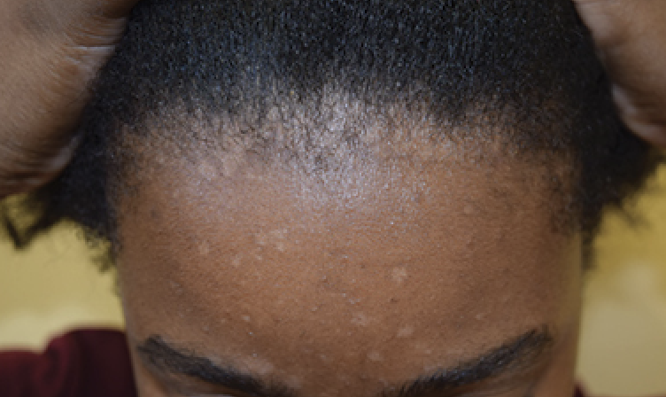
Seborrheic dermatitis may present differently in individuals with skin of color. Darker-skinned individuals may present with scaly, hypopigmented macules and patches in typical areas of involvement. Arcuate or petal-like patches may be seen, specifically termed petaloid seborrheic dermatitis.
Children of color often do not experience the classic “cradle cap” appearance of seborrheic dermatitis, and have erythema, flaking, and hypopigmentation of the affected areas and folds of skin. Seborrheic dermatitis tends to respond well to conventional treatments, although it tends to recur.
Treatment of sebborrheic dermatitis include topical corticosteriods, pimecrolimus cream, salicylic acid, selenium sulfide, etc. Skin of color patients may require a modified treatment approach which takes into account differences in hair texture and hair washing frequency. This paper aims to highlight these differences to help reduce disparities in the management of seborrheic dermatitis in patients of color. J Drugs Dermatol. 2019;18(1):24-27.
Introduction
Scalp and hair disorders are among the most common concerns in patients of color, particularly African-Americans. Seborrheic dermatitis is a chronic, inflammatory condition that affects areas of high sebum production and is a common reason for dermatologic consultations in skin of color patients. A literature exists regarding the presentation and treatment of seborrheic dermatitis in this population. As more African-Americans seek dermatologic care, it becomes crucial for dermatologists to be trained in recognizing and addressing the concerns of diverse patient populations.
This paper addresses the differences in the appearance and treatment of seborrheic dermatitis (SD) in skin of color patients. Highlighting these differences can allow for an effective approach and ultimately reduce the current disparities in the management of skin of color patients with SD.
Epidemiology and Causes
Seborrheic dermatitis (SD) is a common, chronic, benign inflammatory skin disorder of unclear pathophysiology affecting 3% to 12% of the population. SD may have slightly increased incidence among African Americans (6.5%)4 and West Africans (2.9-6%). In a study that compared the most common diagnoses for patients of various ethnic groups in a hospital-based dermatology practice, SD was among the five most common diagnoses observed in black patients.
Seborrheic dermatitis is prominent among black women and can be exacerbated by excessive use of hair oil and pomade, and infrequent shampooing. Some of the recognized risk factors for SD include immunodeficiency (HIV), neurological (Parkinson’s disease) or cardiac disease, as well as alcoholic pancreatitis.
Several factors have been proposed to play a role in the development of SD including the proliferation of the commensal yeast genus, Malassezia, the host immune response, and the composition of sebaceous gland secretions. The population density of Malassezia furfur has been shown to be highest in anatomic sites most highly populated with sebaceous glands, overlapping with areas where SD tends to occur. However, it is unclear if this has a direct impact on the pathogenesis of SD. In 1989, Bergbrant and Faergemann found no difference in the number of yeast cells in patients with SD and healthy controls, but several other studies since that time have suggested a positive correlation between yeast density and severity of SD. A study conducted by Nakabayashi et al compared lesional and non-lesional skin in patients with SD and healthy controls.
For Clinical Manifestations, Treatment Considerations and Prevention, read the free text article.
Get More from the JDD
Get the latest news delivered straight to your Inbox – sign up for the JDD Newsletter.
Discover the latest research, exclusive articles from leading dermatology experts, popular Podcast episodes, free CME activities, and more!


