Featured Article
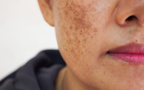
Post inflammatory hyperpigmentation (PIH) is an acquired hyper-melanosis typically arising following inflammatory lesions. It is one of the most common dermatologic complaints, which may develop in all skin types, however, higher prevalence is seen in patients with Fitzpatrick skin types IV-VI.
Credit: Duangjan – stock.adobe.com Copyright: ©Duangjan – stock.adobe.com
Q-Switched 1064 nm Nd:YAG Laser in Treating Axillary Hyperpigmentation in Filipino Women With Skin Types IV-V
Use of a low-fluence 1064 nm Q-switched Nd:YAG laser in the treatment of axillary hyperpigmentation is safe and effective in patients with darker skin type.
Post inflammatory hyperpigmentation (PIH) is an acquired hyper-melanosis typically arising following inflammatory lesions. It is one of the most common dermatologic complaints, which may develop in all skin types, however, higher prevalence is seen in patients with Fitzpatrick skin types IV-VI.
The etiology underlying PIH can either be an exogenous source (allergy, irritation, contact dermatitis, dermabrasion, laser therapy or burns), an endogenous factor (primary inflammatory or bullous dermatosis) or even induced by an infectious agent such as herpes zoster virus infection. Morphologic pattern and degree of pigmentation vary depending on causative factors and melanin distribution in the epidermis, dermis or both.1,2,3
IH typically manifests as macules or patches in the same distribution in previous areas of inflammation, which can be classified as two clinical forms: epidermal and/or dermal. In epidermal PIH, melanocytes are activated, and release melanin resulting in tan brown or dark brown appearance and may take months to years to resolve. Dermal PIH includes activation of basal keratinocytes, which also release melanin, and present as dark brown to blue-grey discoloration that may either be permanent or resolve over an extended period of time if not treated.
Differentiating between the two is difficult, and PIH is probably a result of the combination of both epidermal and dermal lesions.4 The degree of PIH varies depending on the etiological factors, skin type or “chromatic tendency” as well as exposure to UV light, certain medications and cutaneous injuries (trauma or even shaving).1,2,5,6 The discoloration is determined by the distribution and depth of pigment within the skin layers.
The pathogenesis of PIH is often related to an increase in melanin synthesis and/or irregular pigment dispersion resulting from cutaneous inflammation. It is considered the end result of: melanocyte proliferation, melanin synthesis and increased activation of tyrosinase coupled with transfer of melanosomes to neighboring keratinocytes. Although the exact mechanism is not yet fully understood, the rise in melanocyte activity and proliferation has been known to be stimulated by inflammatory mediators such as reactive oxygen species, prostaglandins and leukotrienes.1,5
Axillary hyperpigmentation is a frequent dermatological complaint, characterized by dermal and epidermal PIH mainly associated with women of darker skin types. Etiological theory associates axillary hyperpigmentation of a form of post inflammatory hyperpigmentation due to continuous irritation due to hair removal, cleansing, tight cloths, or innate darkening from genetic related factors.11
The purpose of this case study was to evaluate the efficacy of a 1064nm nanosecond QS Nd:YAG laser in the treatment of axillary PIH in Filipino women with Fitzpatrick skin types IV-V.
You May Also Like