INTRODUCTION
Linear IgA bullous dermatosis (LABD), also known as IgA pemphigoid, is an acquired autoimmune sub-epidermal vesiculobullous disease. Patients with LABD can experience intense pruritus with burning and pain, which may precede relapses of visible lesions.1 Furthermore, LABD can also present with prurigo nodularis-like nodules secondary to chronic itch.2
LABD derives its name from the characteristic linear deposition of IgA and IgG autoantibodies at the basement membrane zone, as seen on direct immunofluorescence. LABD can be idiopathic, drug-induced, or associated with malignancy, inflammatory bowel disease, and other autoimmune diseases.3
LABD mainly affects preschool-aged children and adults, and is a chronic condition, often persisting for years with rare spontaneous remission. In children, lesions typically exhibit a "string of pearls" arrangement, while LABD manifestations in adults are more variable and can appear similar to dermatitis herpetiformis and bullous pemphigoid. Skin lesions can be papular, vesicular, or bullous, and typically have a widespread distribution. Mucosal lesions can also occur, with oral and ocular lesions being the most commonly involved sites.3
Management and treatment of acute onset LABD includes discontinuation of possible offending drugs. First-line treatment is with dapsone, oral corticosteroids, and/or sulfonamides. Additional therapies for recalcitrant disease include Intravenous immunoglobulin (IVIG), colchicine, rituximab, mycophenolate mofetil, and methotrexate.4
We present a case of a patient with LABD who had severe, persistent itch secondary to LABD, with significant improvement on nemolizumab.
Case
A 67-year-old woman with asthma and well-controlled HIV (undetectable viral load) was seen for follow-up of known LABD. The patient was initially diagnosed 4.5 years earlier, with biopsy showing a subepidermal blister with lymphocytic, histiocytic, neutrophilic, and eosinophilic infiltrate as well as perivascular inflammation. Direct immunofluorescence was positive for IgA

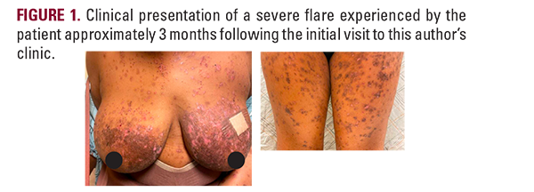
deposition. Deposition of IgG, IgM, and C3 was not observed. The patient had trialed multiple anti-itch treatments with minimal success. Initial management of her itch included gabapentin, which was not well tolerated and discontinued, along with antihistamines, topical steroids, and emollients. Dapsone and colchicine were initiated but discontinued due to dyspnea and gastrointestinal side effects, respectively. The patient also tried pentoxifylline (PTX), which was somewhat helpful, as well as UVB phototherapy, with minimal relief. After the failure of these treatments (Figure 1), the patient sought the authors’ care.
At her first visit to the authors' clinic, the patient had 30% BSA involvement with scattered vesicles/bullae and open erosions on the trunk and extremities as well as erosions on her hard palette (Figure 2). Workup revealed elevated IgE and complement levels with negative ANA and ENA panels. Throughout her treatment, her blood demonstrated eosinophilia. She was started on IV rituximab 375 mg/m² every three months (initiated March 2021) and mycophenolate mofetil 500 mg twice daily (initiated April 2021), which resulted in remission of LABD lesions within 3 months. Her skin and oral lesions continued to improve after initiation of omalizumab in May 2021; however, she experienced rebound pruritus, and the medication was discontinued. Topical ruxolitinib was trialed for itch as well, but was not effective.
IVIG was trialed from August to November 2022 but was discontinued because the patient reported headaches, heartburn, and minimal efficacy. Following IVIG discontinuation, 300 mg dupilumab biweekly was started and the patient reported temporary relief from her pruritus. After 6 months of this treatment, she reported no new lesions but an intensified itch affecting sleep and work. Physical exam was notable for hyperpigmented macules and patches scattered across her


