INTRODUCTION
Pyoderma gangrenosum (PG) is a rare, neutrophilic dermatosis characterized by rapidly enlarging, painful ulcers with violaceous and undermined borders. It is commonly associated with systemic disorders, including rheumatoid arthritis, hematologic malignancies, and inflammatory bowel disease (IBD).1 While PG often correlates with IBD flares, its pathogenesis is not well understood. Colectomy has been used to treat severe PG, suggesting a link between PG and bowel inflammation.2 However, rare reports of PG developing after colectomy and ileostomy, with ulcers at peristomal sites, indicate that PG can occur independently of active bowel disease.3 Here, we present a case of PG that occurred one year after total colectomy for ulcerative colitis (UC). The PG was successfully treated with intralesional triamcinolone and systemic adalimumab. This case supports the observation that PG can manifest independently of bowel disease activity.
Case Presentation
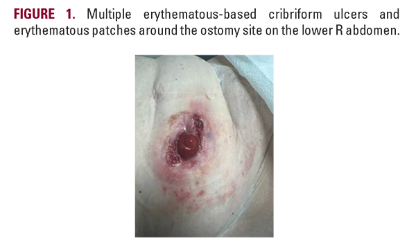
A 55-year-old woman presented with painful ulcerations near her ostomy site one year after undergoing a total colectomy for UC. Her history of UC had been associated with severe refractory diarrhea, fecal incontinence, weight loss, and abdominal pain despite treatment with mesalamine and risankizumab. She also had recurrent Clostridium difficile infections, sepsis requiring fecal transplantation, and a prior sigmoid colectomy for diverticulitis with abscess formation. Due to worsening UC despite medical therapy, she underwent total colectomy and ileostomy. Approximately nine months postoperatively, the patient reported pain and irritation around the ostomy site. She was diagnosed with a chronic intraabdominal abscess and underwent drainage on the medial aspect of the ileostomy. Four months later, the patient developed multiple large cribriform ulcers with undermined borders and surrounding erythema surrounding a clean ostomy (Figure 1). Due to increasing pain, history of UC, undermined borders, cribriform appearance, and progression of ulcerations, peristomal PG was suspected. The patient was initially treated with intralesional triamcinolone injections (10 mg/mL x 1 mL) across two visits. Due to the progression of lesions, systemic adalimumab was initiated (160 mg loading dose, followed by 80 mg, then 40 mg every
Case Presentation
A 55-year-old woman presented with painful ulcerations near her ostomy site one year after undergoing a total colectomy for UC. Her history of UC had been associated with severe refractory diarrhea, fecal incontinence, weight loss, and abdominal pain despite treatment with mesalamine and risankizumab. She also had recurrent Clostridium difficile infections, sepsis requiring fecal transplantation, and a prior sigmoid colectomy for diverticulitis with abscess formation. Due to worsening UC despite medical therapy, she underwent total colectomy and ileostomy. Approximately nine months postoperatively, the patient reported pain and irritation around the ostomy site. She was diagnosed with a chronic intraabdominal abscess and underwent drainage on the medial aspect of the ileostomy. Four months later, the patient developed multiple large cribriform ulcers with undermined borders and surrounding erythema surrounding a clean ostomy (Figure 1). Due to increasing pain, history of UC, undermined borders, cribriform appearance, and progression of ulcerations, peristomal PG was suspected. The patient was initially treated with intralesional triamcinolone injections (10 mg/mL x 1 mL) across two visits. Due to the progression of lesions, systemic adalimumab was initiated (160 mg loading dose, followed by 80 mg, then 40 mg every
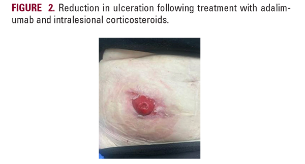
two weeks). Over the following month, her ulcers significantly improved, decreasing from three lesions to a single small ulcer (Figure 2).
DISCUSSION
This case underscores that PG can occur independently of UC disease activity, even after complete removal of the colon. The pathophysiology of PG post-colectomy likely reflects immune dysregulation despite the absence of active bowel disease, including excessive neutrophil recruitment and elevated pro-inflammatory cytokines such as IL-8, IL-1β, TNFα, and IL-36.1,4 Colectomy may also induce intestinal dysbiosis and disrupt immune tolerance by eliminating gut-associated lymphoid tissue, which promotes the production of anti-inflammatory

