INTRODUCTION
Case Synopsis
A 45-year-old woman presented with a one-year history of a growing mass on the scalp. The patient believed the lesion was a benign cyst and requested removal due to its painful nature. Physical exam revealed a 10x24 mm reddish raised nodule on the right parietal scalp initially clinically suspected to be a pilar cyst (Figure 1).
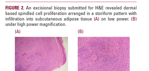
The area appeared inflamed, but without signs of infection. The lesion was removed with excision margins and a tissue sample was sent for histopathological diagnosis. Excisional biopsy of the lesion revealed this to be DFSP extending to the peripheral deep margins. Histological analysis showed dermal based spindled cell proliferation.
No features of fibrosarcomatous transformation were identified. There was additional staining completed for S-100 protein, Melan-A, desmin, and CD34; all of which were negative except for CD34 which stained positive.
The patient’s medical history includes the sickle cell trait, anemia, and migraine headaches. She denied any personal or family history of skin cancers or other cancers, including DFSP. She had no scar or previous trauma history to the lesion site. The patient was further treated with "slow-Mohs" surgery involving 10 mm margins taken around the lesion down to subcutaneous fascia. Focal positivity was found in two sections following stage one and the patient was brought back for a second stage of "slow-Mohs" the next day. An additional layer


