From the Vault: Pulled due to increased interest in rare onychomycosis presentations
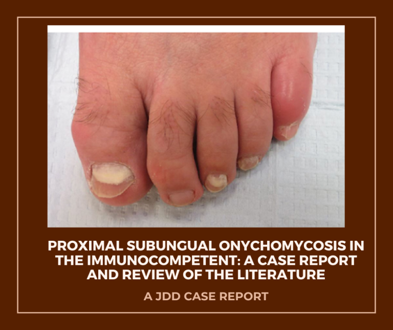
This case report dates back to JDD’s April 2018 issue. It’s a case report describing a healthy 51-year-old man who developed white discoloration and proximal onycholysis of multiple toenails over three months. Examination showed proximal opaque patches, subungual debris, and mild paronychia. Histopathology of proximal nail fragments demonstrated parakeratosis, neutrophilic collections, and hyphae on periodic acid Schiff staining, supporting a diagnosis of proximal subungual onychomycosis with concurrent tinea pedis. The patient improved with oral fluconazole.
Proximal subungual onychomycosis is uncommon outside of immunocompromised states, so this presentation in an apparently healthy host is notable and, as the authors emphasize, should prompt consideration of an evaluation for underlying immunodeficiency. The report is concise and focused on clinical and histologic correlation without extensive technical detail.
For practicing dermatology clinicians and HCPs, this case highlights a diagnostic reminder about nail sampling and pathology when proximal nail changes are present. Read the full case for clinical photos, histology, and the treatment timeline.
J Drugs Dermatol. 2018;17(4):475-478. doi:10.36849/JDD.1888
Blog write-up assisted by AI

