Morphea
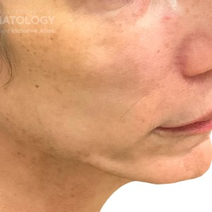
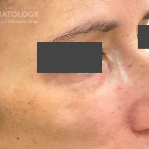
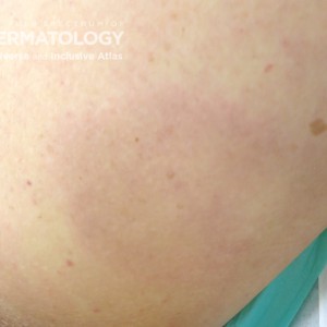
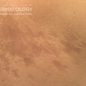
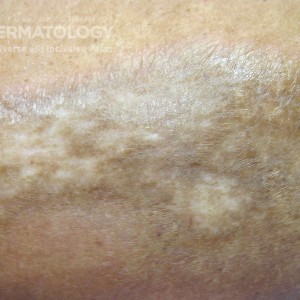
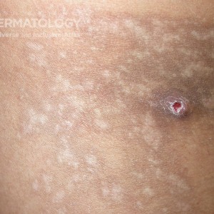
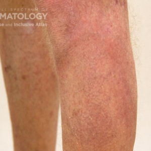
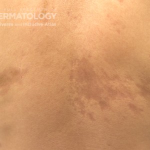
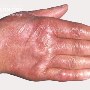
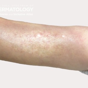
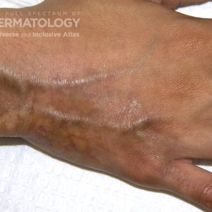
This collection of images displays the various stages of plaque-type morphea across different skin tones, with progression from
the erythematous/edematous phase to hyperpigmented scarred plaques. Notice that in darker skin tones the latter stage is hallmarked by ill-defined central hypopigmentation with peripheral hyperpigmentation. Persistent activity can be discerned by presence of a violaceous peripheral rim to lesions, which is absent in some of the images.
In this collection of photos, active morphea is appreciated in all skin tones. Notable in some of the images: the slate gray hyperpigmentation centrally with peripheral rim of erythema in a lighter skinned patient; subtle atrophy, hallmarked by presence of telangiectasia and mild erythema throughout the lesion extending to 5th digit in a darker toned patient.
Log into your JDD account to access high resolution images and request permissions.

DermAtlas_Morphea_Parry romberg_face-oblique-right-08.30.2023-55633160
https://cms.sanovaworks.com/uploads/2023/10/f9505ee4fffab4089ba96fd2f5dbad1a-small.jpg

DermAtlas_Morphea. Parry romberg_face-oblique-right-08.30.2023-55633160
https://cms.sanovaworks.com/uploads/2023/10/af72bb93a896189a33632b5024df2c6d-small.jpg

morphea type B leg.jpg
https://cms.sanovaworks.com/uploads/2022/09/2e4b98b89d24d6b148bdcb03787be8dd-small.jpg

morphea_b_carrington_back2.jpg
https://cms.sanovaworks.com/uploads/2022/09/542cf9222f28df4a9eefa1cb95efdd47-small.jpg

morphea type D abdomen.jpg
https://cms.sanovaworks.com/uploads/2022/09/a83c29c6b74e52f6a6c7a5d9b01c66db-small.jpg

Morphea type D .jpg
https://cms.sanovaworks.com/uploads/2022/09/0b60e11ed5fe78d4aca384e12f3aedff-small.jpg

Morphea_A_Adusumilli_Leg1.jpg
https://cms.sanovaworks.com/uploads/2022/09/ee6b9bb93a151e612efff8e633749fc2-small.jpg

morphea_b_carrington_fullback2.jpg
https://cms.sanovaworks.com/uploads/2022/09/e0641459a4af63382c02027fee444353-small.jpg

Morphea_DIR_58358_.jpg
https://cms.sanovaworks.com/uploads/2022/09/20dba76be0954869acba07b477fd8722-small.jpg

morphea type B arm23_.jpg
https://cms.sanovaworks.com/uploads/2022/09/4fcf2f0af55f24ff920e7f5873aa7f4e-small.jpg

morphea_c_carrington_hand2.jpg
https://cms.sanovaworks.com/uploads/2022/09/0374b4f79a75824b93435af299f320e1-small.jpg


