Herpes Zoster
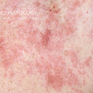
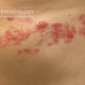
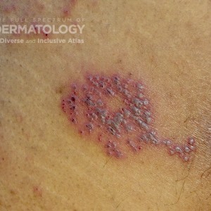
                            This collection of images illustrate the dermatomal distribution of grouped, umbilicated vesicles (often with gray, dusky fluid) which 
is a helpful feature when diagnosing herpes zoster; however, keep in mind that erythematous papules and vesicles might be challenging to recognize during the earlier stages of the clinical presentation.
Log into your JDD account to access high resolution images and request permissions.


                        JDD_C1485_Acloseup.jpg
                        
                        
                        https://cms.sanovaworks.com/uploads/2022/09/e817b64d27db3d2b81a462a89c49fd55-small.jpg


                        HerpesZoster_A_1.jpg
                        
                        
                        https://cms.sanovaworks.com/uploads/2022/09/21dde6cff29b504231f373fe0b36c391-small.jpg


                        Herpes_Zoster_C_2.jpg
                        
                        
                        https://cms.sanovaworks.com/uploads/2022/09/666ac14775e58720debff387c0a1ba4d-small.jpg