INTRODUCTION
Eyebrows play an essential role in facial symmetry, communication, and aesthetic identity.1,2 Scarring, or cicatricial, eyebrow alopecia involves a diverse group of inflammatory, autoimmune, infectious, and traumatic processes that destroy the follicular stem cell and eliminate the potential for regrowth.3,4 These disorders can be diagnostically challenging, often requiring careful clinical evaluation with trichoscopy to distinguish them from nonscarring etiologies.4 Because delayed recognition may lead to irreversible loss and significant psychosocial distress, timely identification and intervention are critical.3,5 Part two summarizes the major scarring causes of eyebrow alopecia and outlines current therapeutic approaches
MATERIALS AND METHODS
A comprehensive literature review was conducted to evaluate the clinical evidence related to the causes, diagnostic features, and management of eyebrow madarosis. Studies were identified using PubMed searches performed in August 2025 (1960–2025) with keywords including "eyebrow alopecia," "eyebrow madarosis," "eyebrow loss," "alopecia areata eyebrow," "frontal fibrosing alopecia eyebrow," "eyebrow hypothyroidism," "eyebrow chemotherapy alopecia," and other etiology-specific terms. Articles were included if they met the following criteria: English-language or English translation available; original studies (including case reports, case series, retrospective or prospective reviews, or clinical trials); and explicit evaluation of eyebrow involvement in alopecia or systemic disease. Studies were excluded if they were review articles without new patient data, expert opinions or commentaries, non-English without translation, or focused exclusively on scalp alopecia without eyebrow-specific outcomes. Each study was assessed for details regarding etiology of eyebrow alopecia, diagnostic methods, interventions, and treatment outcomes. For Part II, analysis was limited to scarring causes of eyebrow alopecia, and etiologies were further subcategorized by underlying disease process.
RESULTS AND DISCUSSION
Scarring Eyebrow Alopecia Autoimmune
Frontal Fibrosing Alopecia Frontal fibrosing alopecia (FFA) is a cicatricial alopecia characterized by progressive frontal hairline recession, with
Frontal Fibrosing Alopecia Frontal fibrosing alopecia (FFA) is a cicatricial alopecia characterized by progressive frontal hairline recession, with
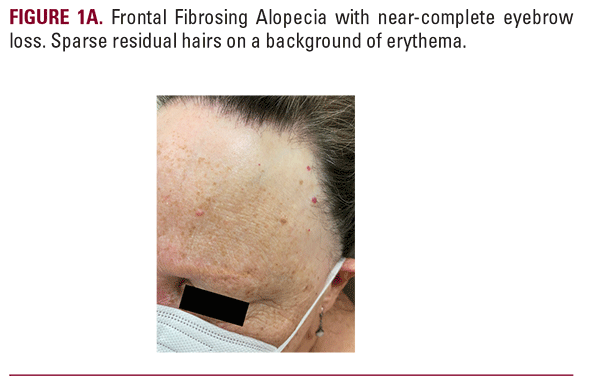
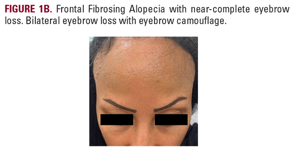
eyebrow involvement reported in up to 96% of cases (Figure 1A and Figure 1B).6,7 Eyebrow loss may even precede scalp involvement, serving as an early clinical sign of disease.8 Trichoscopy typically reveals tapered or broken hairs, pinpoint white dots, vellus hairs, and dystrophic hairs.9 In contrast to scalp FFA, affected eyebrows can demonstrate nonscarring features on dermoscopy and histology, including preservation of follicular ostia and sebaceous glands.10
Management of FFA includes anti-inflammatory therapies aimed at slowing disease progression and providing symptom relief. Treatment options include topical agents (highpotency corticosteroids, calcineurin inhibitors, intralesional triamcinolone, and platelet-rich plasma therapy) and systemic agents such as corticosteroids, hydroxychloroquine, mycophenolate, retinoids, and 5-α-reductase inhibitors (dutasteride and finasteride), which have shown benefit.11 Initial management typically includes 5-alpha reductase inhibitors (dutasteride 0.5 mg/day or finasteride 1–5 mg/day) with hair loss stabilization in 88% (158/180) of FFA patients.12 Newer therapies, including topical and oral JAK inhibitors (tofacitinib, ruxolitinib, delgocitinib, baricitinib, and brepocitinib), have shown encouraging efficacy in reducing inflammation, stabilizing disease progression, and improving clinical severity scores.13 Biologics, such as tildrakizumab, have also shown promise in refractory cases, while adjunct therapies like laser/light devices have also been considered.13
Although no standardized treatment exists for eyebrow management, several therapeutic approaches have been reported. Intralesional triamcinolone acetonide (ILTAC) at a concentration of 10 mg/mL, with 0.125 mL injected per eyebrow, resulted in regrowth in 10 of 11 patients with partial eyebrow loss at 2- to 6-month follow-up.14 In clinical practice, ILTAC concentrations for eyebrow involvement vary; while 10 mg/mL has been reported in the literature, many providers use lower concentrations (2.5–5 mg/mL) to mitigate the risk of localized cutaneous atrophy. Oral dutasteride 0.5 mg and finasteride 2.5 mg have been described in case reports and small series, with some improvement noted.15 Hair transplantation may be considered after inflammatory processes have stabilized, though outcomes in patients with FFA can vary. Hair transplantation has been attempted, with eight out of ten patients initially demonstrating hair growth and satisfactory cosmetic results; however, most experienced loss of transplanted hairs after three to four years, with only one patient maintaining durable regrowth.16 Prostaglandin analogs such as bimatoprost have been reported to promote eyebrow regrowth in two out of three patients when used twice daily for nine months.17,18 In another report, seven women with biopsy-confirmed FFA and moderate eyebrow loss were treated with low-dose oral minoxidil (0.5–1.25 mg/day, titrated to 2.5 mg/day in most); and after 6 months, all achieved cosmetically acceptable regrowth, with partial response in five and near-complete response in two.19 Ultimately, several therapeutic approaches have shown potential benefit for eyebrow involvement in FFA. Oral and topical formulations of JAK inhibitors, which have demonstrated efficacy for scalp stabilization, suggest a promising therapeutic option for eyebrow management.
Localized Scleroderma (En Coup de Sabre)
Localized scleroderma, particularly the linear morphea subtype known as en coup de sabre, may extend across the forehead and scalp and can result in eyebrow atrophy. In a review of 50 pediatric patients with linear morphea, eyebrow loss was reported in approximately 4% of cases.20,21 Treatment aims to control inflammation and prevent progression through topical, phototherapeutic, and systemic approaches. Topical corticosteroids, intralesional triamcinolone, calcipotriene, and tacrolimus are suitable for limited or superficial lesions.22 Phototherapy with narrowband UVB is effective for widespread superficial disease, while broadband UVA or UVA1 is preferred for deeper dermal involvement.22 Systemic therapy with methotrexate, corticosteroids, or both is indicated for deep, disabling, or rapidly progressive cases. Methotrexate (15–25 mg/ week) with corticosteroids (prednisone 0.5–1 mg/kg daily or intravenous methylprednisolone 30 mg/kg/day) remains first-line; mycophenolate mofetil (500–2500 mg daily), hydroxychloroquine, or phototherapy may be considered if refractory.22,23 In a double-blind randomized controlled trial with 70 pediatric morphea patients, oral methotrexate (15 mg/
Management of FFA includes anti-inflammatory therapies aimed at slowing disease progression and providing symptom relief. Treatment options include topical agents (highpotency corticosteroids, calcineurin inhibitors, intralesional triamcinolone, and platelet-rich plasma therapy) and systemic agents such as corticosteroids, hydroxychloroquine, mycophenolate, retinoids, and 5-α-reductase inhibitors (dutasteride and finasteride), which have shown benefit.11 Initial management typically includes 5-alpha reductase inhibitors (dutasteride 0.5 mg/day or finasteride 1–5 mg/day) with hair loss stabilization in 88% (158/180) of FFA patients.12 Newer therapies, including topical and oral JAK inhibitors (tofacitinib, ruxolitinib, delgocitinib, baricitinib, and brepocitinib), have shown encouraging efficacy in reducing inflammation, stabilizing disease progression, and improving clinical severity scores.13 Biologics, such as tildrakizumab, have also shown promise in refractory cases, while adjunct therapies like laser/light devices have also been considered.13
Although no standardized treatment exists for eyebrow management, several therapeutic approaches have been reported. Intralesional triamcinolone acetonide (ILTAC) at a concentration of 10 mg/mL, with 0.125 mL injected per eyebrow, resulted in regrowth in 10 of 11 patients with partial eyebrow loss at 2- to 6-month follow-up.14 In clinical practice, ILTAC concentrations for eyebrow involvement vary; while 10 mg/mL has been reported in the literature, many providers use lower concentrations (2.5–5 mg/mL) to mitigate the risk of localized cutaneous atrophy. Oral dutasteride 0.5 mg and finasteride 2.5 mg have been described in case reports and small series, with some improvement noted.15 Hair transplantation may be considered after inflammatory processes have stabilized, though outcomes in patients with FFA can vary. Hair transplantation has been attempted, with eight out of ten patients initially demonstrating hair growth and satisfactory cosmetic results; however, most experienced loss of transplanted hairs after three to four years, with only one patient maintaining durable regrowth.16 Prostaglandin analogs such as bimatoprost have been reported to promote eyebrow regrowth in two out of three patients when used twice daily for nine months.17,18 In another report, seven women with biopsy-confirmed FFA and moderate eyebrow loss were treated with low-dose oral minoxidil (0.5–1.25 mg/day, titrated to 2.5 mg/day in most); and after 6 months, all achieved cosmetically acceptable regrowth, with partial response in five and near-complete response in two.19 Ultimately, several therapeutic approaches have shown potential benefit for eyebrow involvement in FFA. Oral and topical formulations of JAK inhibitors, which have demonstrated efficacy for scalp stabilization, suggest a promising therapeutic option for eyebrow management.
Localized Scleroderma (En Coup de Sabre)
Localized scleroderma, particularly the linear morphea subtype known as en coup de sabre, may extend across the forehead and scalp and can result in eyebrow atrophy. In a review of 50 pediatric patients with linear morphea, eyebrow loss was reported in approximately 4% of cases.20,21 Treatment aims to control inflammation and prevent progression through topical, phototherapeutic, and systemic approaches. Topical corticosteroids, intralesional triamcinolone, calcipotriene, and tacrolimus are suitable for limited or superficial lesions.22 Phototherapy with narrowband UVB is effective for widespread superficial disease, while broadband UVA or UVA1 is preferred for deeper dermal involvement.22 Systemic therapy with methotrexate, corticosteroids, or both is indicated for deep, disabling, or rapidly progressive cases. Methotrexate (15–25 mg/ week) with corticosteroids (prednisone 0.5–1 mg/kg daily or intravenous methylprednisolone 30 mg/kg/day) remains first-line; mycophenolate mofetil (500–2500 mg daily), hydroxychloroquine, or phototherapy may be considered if refractory.22,23 In a double-blind randomized controlled trial with 70 pediatric morphea patients, oral methotrexate (15 mg/