INTRODUCTION
Herpes zoster (HZ), caused by the reactivation of the varicella-zoster virus, typically resolves without long-term sequelae. However, chronic dermatologic complications can emerge at the site of prior infection, a phenomenon known as an isotopic response.1 A wide spectrum of dermatologic conditions has been reported with isotopic response, highlighting its diverse clinical manifestations. Herein, we present 2 cases of bullous pemphigoid and discoid lupus erythematosus to underscore this variability and expand awareness of this phenomenon.
Case 1
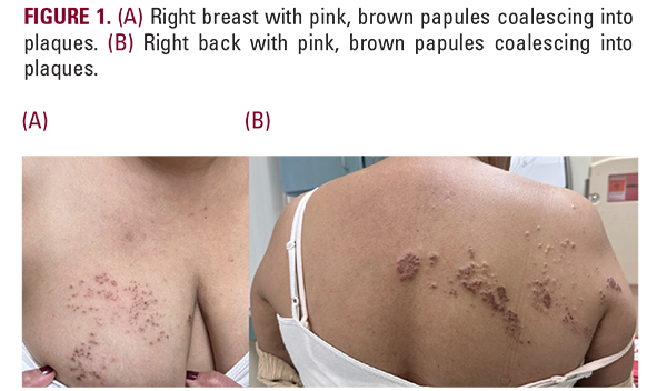
A 51-year-old woman presented with a six-month history of a persistent rash following an episode of HZ. Her medical history was notable for SLE with lupus nephritis, hypothyroidism, and psoriasis vulgaris. During her HZ episode, she was treated with antiviral therapy, but her SLE medications, prednisone and mycophenolate mofetil (MMF), were held. She remained on hydroxychloroquine 400 mg daily. The patient endorsed chronic dry eyes and intermittent stabbing pain in the affected skin areas. On examination, the rash consisted of multiple pink, brown papules coalescing into plaques overlying the midline of the back extending anteriorly to the right breast in T1-T4 dermatome (Figure 1). A skin biopsy revealed basal vacuolar alteration, and superficial and deep perivascular and periadnexal lymphocytic infiltrate. Viral cytopathic changes were absent, supporting the diagnosis of DLE rather than
Case 1
A 51-year-old woman presented with a six-month history of a persistent rash following an episode of HZ. Her medical history was notable for SLE with lupus nephritis, hypothyroidism, and psoriasis vulgaris. During her HZ episode, she was treated with antiviral therapy, but her SLE medications, prednisone and mycophenolate mofetil (MMF), were held. She remained on hydroxychloroquine 400 mg daily. The patient endorsed chronic dry eyes and intermittent stabbing pain in the affected skin areas. On examination, the rash consisted of multiple pink, brown papules coalescing into plaques overlying the midline of the back extending anteriorly to the right breast in T1-T4 dermatome (Figure 1). A skin biopsy revealed basal vacuolar alteration, and superficial and deep perivascular and periadnexal lymphocytic infiltrate. Viral cytopathic changes were absent, supporting the diagnosis of DLE rather than