INTRODUCTION
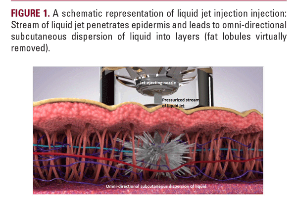
Jet injection provides an intradermal drug delivery through invasive but needle-free route of administration. The modality implements a pressurized stream of liquid that penetrates the skin with a minimal injury to epidermis and dermis. The principal mode of action is based on the synergy between injected drug and the micro-trauma generated in the soft tissues by the dispersion. Once penetrated, the micro-droplets of the injected drug are dispersed in multiple 3-D directions increasing the area and contact between the skin and the drug (Figure 1). Kinetic energy of the jet powers the droplets to travel and originate a micro-trauma impact on the surrounding tissue. Extent of the spread is limited by the friction with surrounding skin and is influenced by heterogeneity and rigidity of the treated region.1
The modality steadily gains popularity in aesthetic dermatology for correction of various skin conditions. Clinical use of the jet technology targets different anatomical structures that require absolute precision in the drug delivery: superficial dermis for correction of the age-related dermal atrophy,2,3,4 dermo-subcutaneous junction for remodeling of atrophic scars,5,6,7 or deep reticular dermis for skin laxity.8
Experiments on the skin-mimicking phantom and porcine skin verified that penetration is a function of the velocity and diameter of jet stream.9,10 It was clinically revealed that injection pressure is directly related to the jet’s depth of penetration8 and controls the level at which dispersion of the injected drug occurs (Figure 2). The ejection volume of the drug and the skin properties was histologically shown to have a direct effect on the side-wise distribution of the injected material.1
As the injection pressure and ejection volume are two parameters that can contribute to the treatment effectiveness, we conducted a series of anatomical experiments in order to investigate further the ability of jet-injection to deliver liquid materials into various dermal and subcutaneous structures.


MATERIALS AND METHODS
At the Anatomical Institute of the University of Graz, two heads conserved with Thiel's solution were used to calculate experimental applications.
Substances, prescribed for clinical applications (NaCl, 20 mg/ml cross-linked Restylane hyaluronic acid, 20% glucose) were used as injection substances and mixed with anatomical color markers (Pintasol E-WL5 blue, E-WL41 oxide red, E-WL61 oxide green (Figure 3a). The dyes were added to mark the
Substances, prescribed for clinical applications (NaCl, 20 mg/ml cross-linked Restylane hyaluronic acid, 20% glucose) were used as injection substances and mixed with anatomical color markers (Pintasol E-WL5 blue, E-WL41 oxide red, E-WL61 oxide green (Figure 3a). The dyes were added to mark the


