DISCUSSION
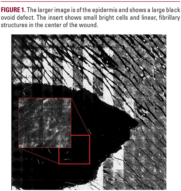
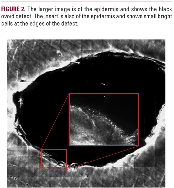
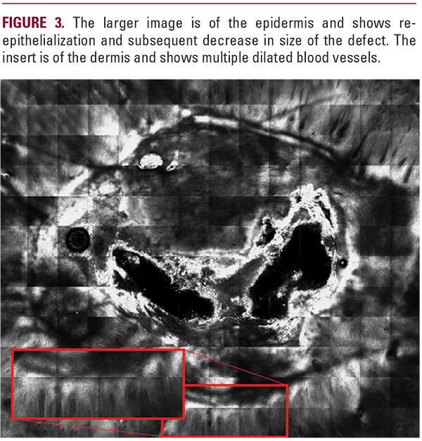
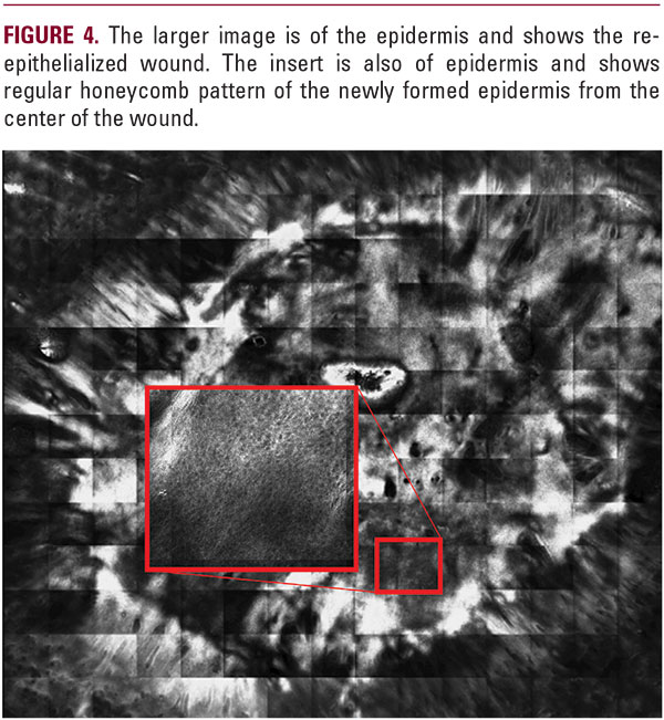
The RCM images on day 1 show small bright cells and fibrillary structures that may correspond to neutrophils and exudate of the inflammatory phase. The RCM images on day 7 and 21 show an irregular honeycomb pattern and decrease in defect size, corresponding to re-epithelialization. The RCM image on day 21 shows multiple dilated vessels, corresponding to new vessel growth of the proliferative phase of wound healing. The RCM image on day 28 shows a regular honeycomb pattern and minimal small bright cells corresponding to resolving inflammation and the tissue remodeling phase of wound healing. RCM imag

