
tion while on chemotherapy. MSH increases cyclic adenosine monophosphate and tyrosine within melanocytes; subsequently,
these elevated levels of tyrosine may stimulate the increased
production of melanin.3
Interferon-α is used in the treatment of chronic myelogenous leukemia. It is also used in the treatment of hepatitis C in combination with ribavirin. It has been postulated that it has the same mechanism of action as doxorubicin, increasing the expression of MSH. It cannot be excluded that ribavirin, which was
concurrently used in the patients being treated for hepatitis C, contributed to the development of lingual hyperpigmentation.4
The lunula is the moon-shaped visible white portion of the
distal nail matrix. Blue lunula is more readily observed on the
digits of the hand and most readily observed on the thumbs.
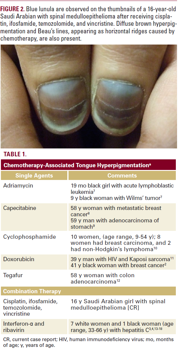
Blue lunula has been caused by congenital disorders, drugs,
heavy metals, and as an idiopathic occurrence (Tables 2 and 3).
Drug-associated blue lunula has been observed not only after
the administration of antineoplastic agents, but also after
treatment with other medications such as minocycline, phenolphthalein, and zidovudine (Table 3). Similar to our patient, individual reports describe patients who developed blue lunula while receiving treatment with combination chemotherapy. To the best of our knowledge, none of these patients also had hyperpigmentation of the tongue.

